Ad Talk to One of Our Highly Flexible Tissue Arrays. DXA is todays established standard for measuring bone mineral density BMD.
 Dexa Dual Energy X Ray Absorptiometry Youtube
Dual Energy X-Ray Absorptiometry DXADEXA is a technique used to assess body composition providing measurements of bone mineral density and content.

Dual energy x ray absorptiometry dxa. Although studies in sedentary populations have investigated the validity of DXA assessment of. Dual Energy X-ray Absorptiometry1 or DEXA or bone densitometry is used primarily for osteoporosis 2tests. Ad Check Price and Availability from a Qpled Supplier Contact a Product Specialist.
Increases Bone Strength Builds Bone Density Stimulates Bone Growth. Ad Clinically proven to increase your height naturally. Its also known as a DXA dual X-ray absorptiometry a bone density scan or a bone densitometry scan Bone density scan DEXA scan - NHS.
Overview of Dual Energy X-Ray Absorptiometry DXA will be used to assess overall skeletal changes that often occur with age by measuring bone mineral content BMC and bone mineral density BMD. Dual-energy X-ray absorptiometry DXA is a two-dimensional imaging technology developed to assess bone mineral density BMD of the entire human skeleton and also specifically of skeletal sites known to be most vulnerable to fracture. The clinical utility of DXA is highly dependent on the quality of the scan acquisition analysis and interpretation.
Ad Clinically proven to increase your height naturally. Dual-energy x-ray absorptiometry DEXA or DXA is a technique used to aid in the diagnosis of osteopenia and osteoporosis. Increases Bone Strength Builds Bone Density Stimulates Bone Growth.
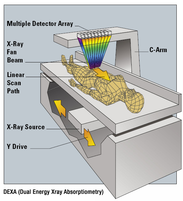
Furthermore with regard to dual energy X-rays absorptiometry DXA there may be important differences between the measures of regions of interest ROI automatically performed by DXA or manually by an evaluator which can cause measurement error and influence the evaluation or diagnosis. Two narrow x-ray beams are emitted at a 90 degree angles across the patient. The most commonly imaged areas are he hip head of the femur lower back lumbar spine or heel calcaneum One peak is absorbed by soft tissue and the other by bone.
Ad Check Price and Availability from a Qpled Supplier Contact a Product Specialist. The most widely used is a scan called dual energy X-ray absorptiometry DXA or DEXA. The test determines bone health and your risk of fracture due to.
Radiographic features Values are calculated for the lumbar vertebrae and femur preferentially and if one of those sites. Dual energy X-ray absorptiometry DXA is rapidly becoming more accessible and popular as a technique to monitor body composition especially in athletic populations. Bone density scanning also called dual-energy x-ray absorptiometry DXA or bone densitometry is an enhanced form of x-ray technology that is used to measure bone loss.
Along with lean and fat mass measurements. In essence each pixel on the x-ray image is assigned to one of three categories bone fat or lean mass. Ad Talk to One of Our Highly Flexible Tissue Arrays.
A DEXA scan is a special type of X-ray that measures bone mineral density BMD. DXA measurements can also be used to provide information on early gender and ethnic changes in the rate of bone accretion and to determine the. Dual-energy X-ray absorptiometry DXA is a technology that is widely used to diagnose osteoporosis assess fracture risk and monitor changes in bone mineral density BMD.
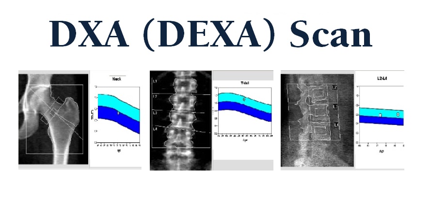
 A Bone Dual Energy X Ray Absorptiometry Dxa Scan For Patient 2 Date Download Scientific Diagram
 How Are Dual Energy X Ray Absorptiometry Dxa Dexa Scans Affected By Surface Stability Physics Stack Exchange
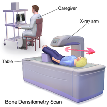
 Dexa Scan Department Of Radiology Uc Davis Health

Dual Energy X Ray Absorptiometry
 The Pitfalls Of Body Fat Measurement Part 6 Dual Energy X Ray Absorptiometry Dexa Weightology
 Dual Energy X Ray Absorptiometry Wikipedia
 Amandeep Hospital Introduces Dexa Scan In Amritsar
 Sample Dual Energy X Ray Absorptiometry Dxa Bone And Tissue Images Download Scientific Diagram
 Absorptimetri Wikipedia Bahasa Indonesia Ensiklopedia Bebas
 Dual Energy X Ray Absorptiometry Dxa Of Osteoporosis Lebanon County Pennsylvania
 Figure 1 From Methodology Review Using Dual Energy X Ray Absorptiometry Dxa For The Assessment Of Body Composition In Athletes And Active People Semantic Scholar
 E Initial Dual Energy X Ray Absorptiometry Dxa Report Showing A The Download Scientific Diagram
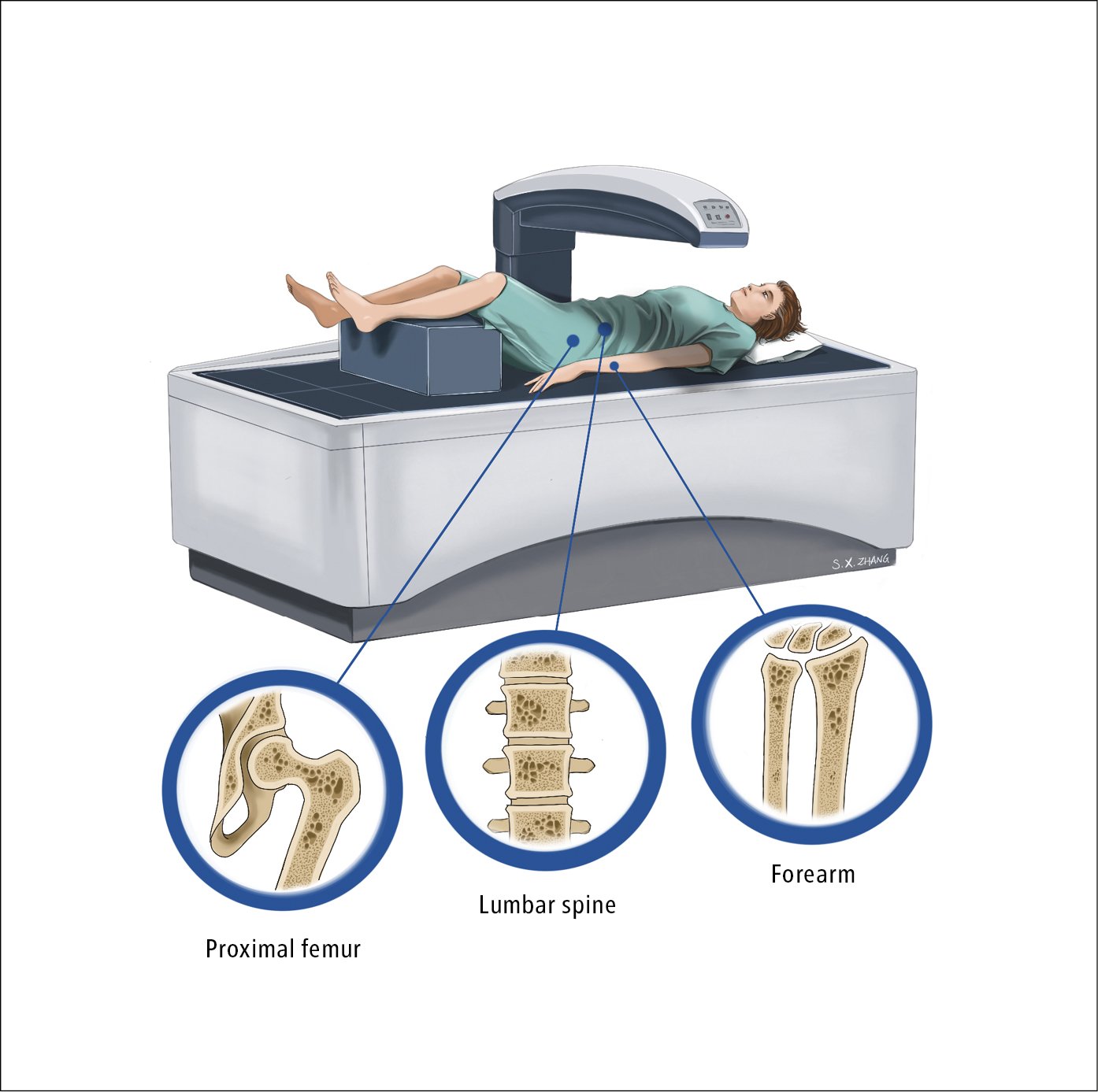
 Figure 031 4288 Dual Energy X Ray Absorptiometry Dxa Showing The Proximal Femur Lumbar Spine And Bones Of The Forearm Illustration Courtesy Of Dr Shannon Zhang Mcmaster Textbook Of Internal Medicine
No comments:
Post a Comment
Note: Only a member of this blog may post a comment.